Sialendoscopy Method (Salivary Gland Endoscopy)

What is Sialendoscopy?

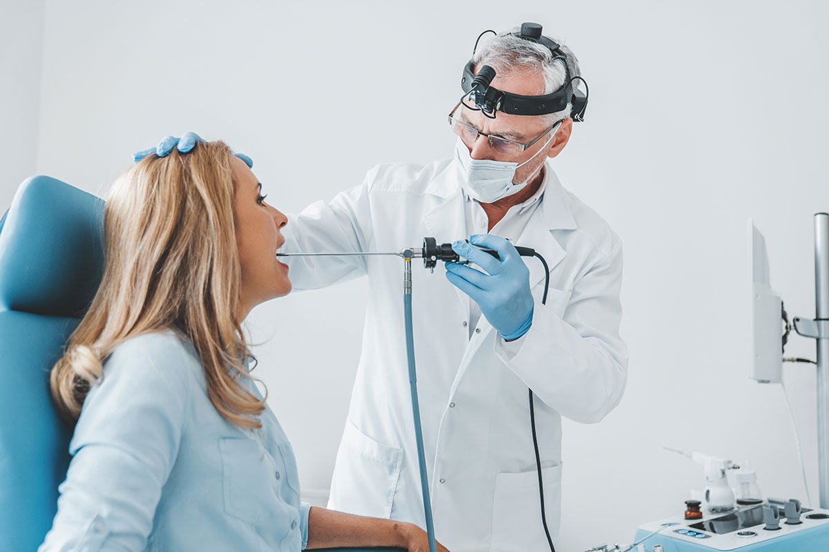
Sialendoscopy is an endoscopic procedure used to visualize the salivary gland ducts and diagnose and treat salivary gland disorders. Initially, the pathology inside the duct is diagnosed, and then endoscopic treatment is applied either during the same session or in a subsequent session.

Sialendoscopy

In which salivary gland disorders is sialendoscopy used?

Salivary gland disorders can be categorized into "obstructive" and "non-obstructive" types. Chronic diseases and infections of the salivary glands occur. Additionally, in conditions like radioactive iodine treatment or certain autoimmune diseases, reduced salivary gland function or gland swelling can be observed. Salivary gland duct obstructions, such as stones or strictures, can also lead to decreased saliva production, resulting in dry mouth. Sialendoscopy is beneficial in diagnosing and treating infections causing recurrent salivary gland swelling and all these health issues.

How does the sialendoscopy procedure work, and how long does it take?

With telescopes of 1.6 or 1.1 millimeters in diameter, access is gained to the salivary gland ducts through the oral cavity and visualized. The telescopes enter through the tiny openings of the ducts, and the ducts are expanded with the help of telescopes, allowing observation throughout the entire duct. Issues such as stones or strictures in the duct can be treated in this way. The procedure duration can vary; it may take up to 1 hour, but in some cases, it can extend to 5-6 hours. This is because, in some situations, large stones may be encountered, requiring breaking and removal, which can be time-consuming. Stones can sometimes be removed directly, or they may be broken into smaller pieces using lasers or lithotripsy, and then extracted in one or more sessions. The factors affecting the process vary from patient to patient and from disease to disease.

What are the advantages of sialendoscopy for patients?

Previously, treating these disorders required major surgeries, but with this new endoscopic system, treatments have become more non-invasive. Consequently, complications and procedure times have decreased, hospital stay durations have been reduced, and the overall comfort of the treatment process has improved for patients. Sometimes, sialendoscopy may need to be combined with more limited oral surgeries, especially open surgeries.

Can the procedure be applied to every patient?

Due to anatomical reasons, it may not be possible to access the salivary gland ducts through the oral cavity for some patients. Sometimes, the ducts may be too narrow or problematic. Apart from such exceptional cases, this method can generally be applied to everyone.

Is there any pain or discomfort during the sialendoscopy procedure? Is anesthesia required?

Both local and general anesthesia can be used. If general anesthesia is chosen, the patient needs to stay under observation for one night. If local anesthesia is preferred, the patient can walk comfortably home at the end of the day after the procedure.

Is there any special preparation required before the sialendoscopy procedure?

No special preparation is required for this procedure. The patient is only asked to prepare for anesthesia, and information is provided about this. Typically, ultrasound and tomography are needed as diagnostic tests. These imaging methods are used to view the condition of the salivary gland, locate stones if present, or show any strictures.

Is there a difference in success rates when comparing sialendoscopy to open surgery?

In open surgery, the salivary gland is generally completely removed. This major and somewhat radical surgical operation naturally carries more risks. In contrast, sialendoscopy is a less invasive procedure performed without removing the salivary gland and largely preserving it.

How widespread is the use of sialendoscopy?

The use of sialendoscopy is increasing over time. The method has become popular worldwide, especially in the last 10 years. There are 1-2 international associations dedicated to sialendoscopy. In Turkey, it is used in approximately 10 centers, including some university and training hospitals, private hospitals, and clinics. This device was first used in Turkey around 2008-2009. As of 2024, Anadolu Health Center is one of the few centers in Turkey offering this procedure. The reason for its limited spread is its cost and complexity, a situation not unique to Turkey but also seen globally.

When should one definitely consult a doctor?

If symptoms such as dry mouth due to reduced saliva production, swelling of the salivary gland during eating, recurrent salivary gland infections and swellings, reduced saliva after radioactive iodine treatment, difficulty swallowing, or an increase in various dental infections are observed, one should consult an ENT specialist.

Medical Technologies

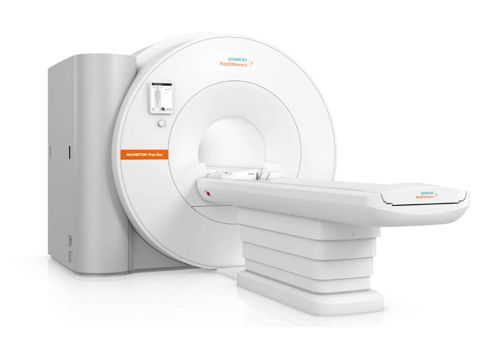
All Technologies
Fibroscan

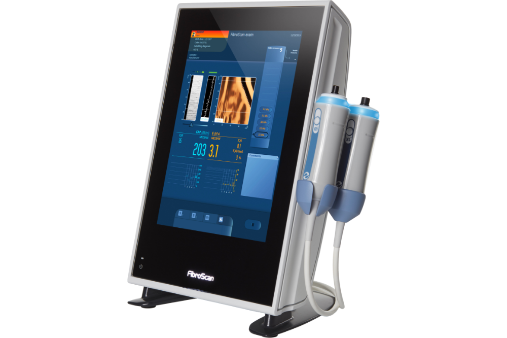
The gold standard in the assessment of liver diseases is liver biopsy. However, thanks to the newly-devised Fibroscan technology, ultrasound waves are now used instead of biopsies in many liver diseases.

More information
Fibroscan
Hybrid Operating Room

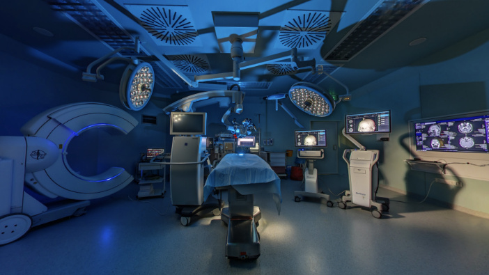
"Hybrid operating room" refers to a new concept of operating rooms where advanced medical imaging systems and medical devices can be used simultaneously.

More information
Hybrid Operating Room
Robotic Surgery

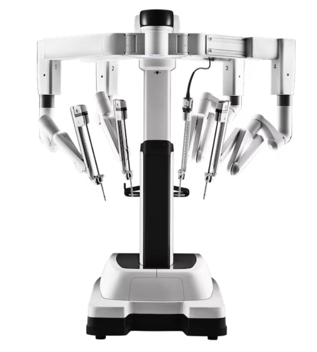
Robotic surgery or robot-assisted surgery, "da Vinci" system of small holes can be defined as the realization of some operations.

More information
Robotic Surgery
CyberKnife M6

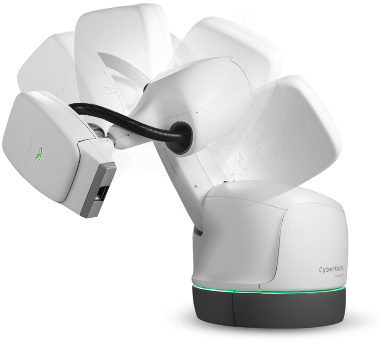
Eliminate tumor surgically lifting up a certain limit, it is a system that prevents the harm of radiation to normal tissues.

More information
CyberKnife M6
Varian Edge

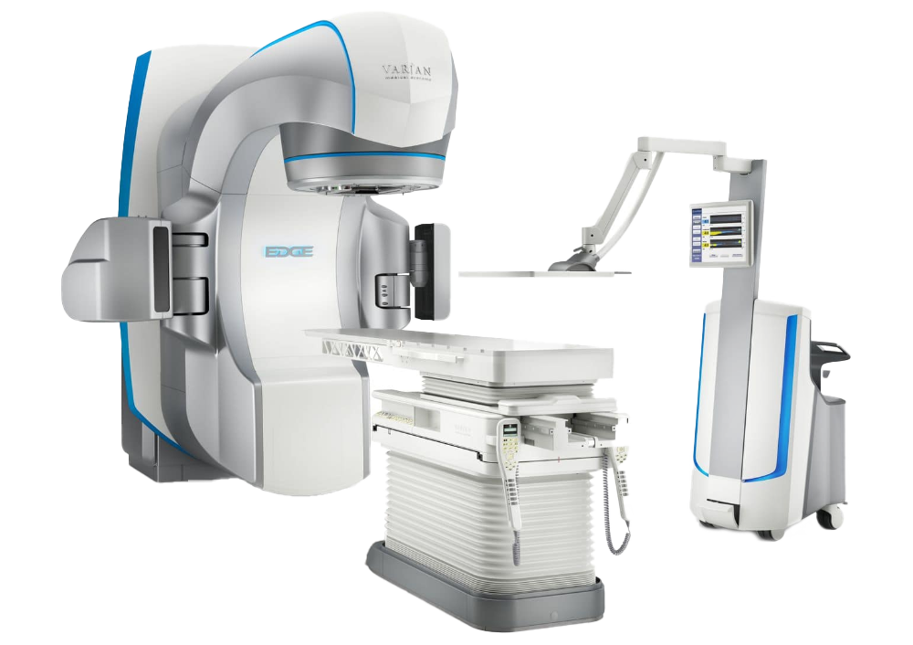
The Varian Edge, first used in Turkey at Anadolu Sağlık Merkezi, is one of the advanced radiotherapy devices that combines the most advanced technologies in cancer radiotherapy.

More information
Varian Edge
Radixact

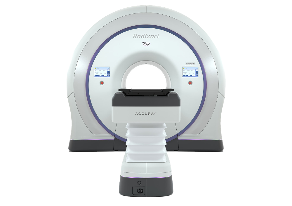
The Radixact device, produced by Accuray, is the newest model in the "tomotherapy" device family.

More information
Radixact
PET-CT

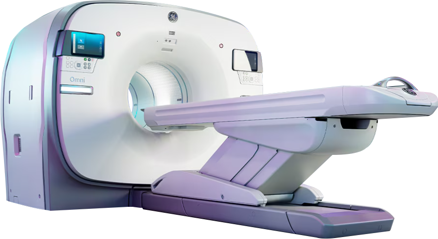
PET/CT, PET (Positron Emission Tomography) and CT (Computed Tomography) techniques combining in the same device is an imaging method that provides more information than they give them separately.

More information
PET-CT
Intraoperative Radiotherapy

In the treatment of breast cancer, radiotherapy today focuses on delivering lower intensity, lower doses, to fewer areas, and for shorter durations.

More information
Intraoperative Radiotherapy
MR-TRUS Fusion

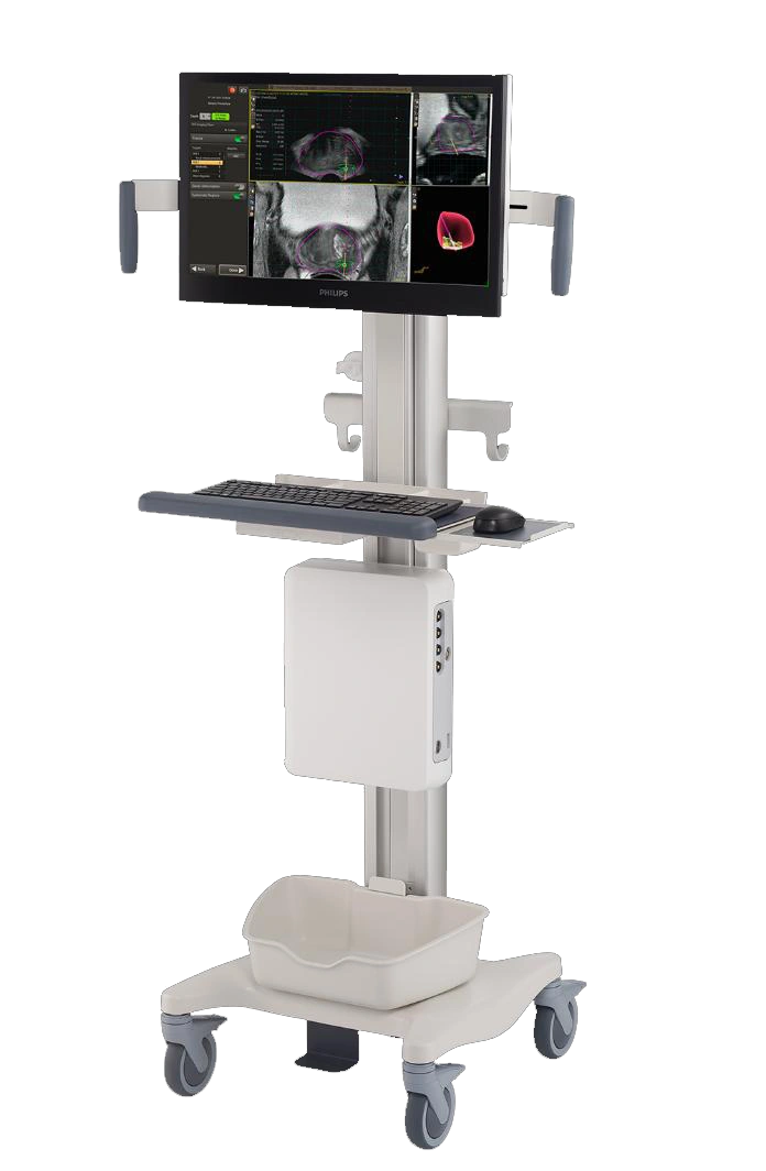
Tumor targeted MR-TRUS Fusion Biopsy is a method, which detects foci suspicious of prostate cancer, and ensures taking biopsy from these foci with millimetric sensitivity by means of an advanced technology device and software.

More information
MR-TRUS Fusion
Radiology Equipment

Radiology Equipment

More information
Radiology Equipment
Ultrasonography

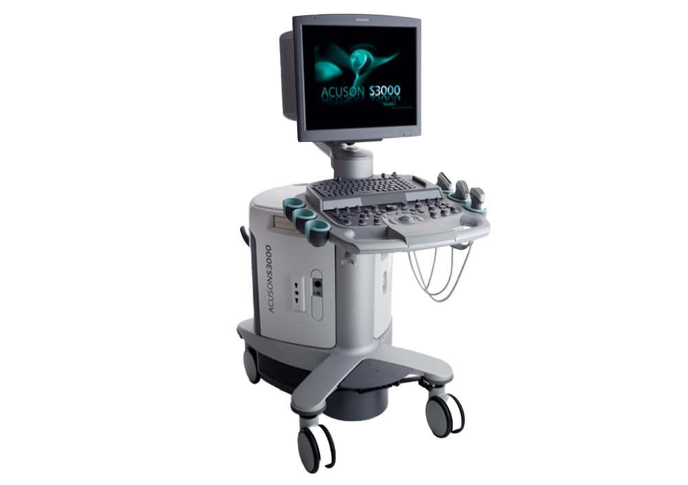
Ultrasound, or more formally, ultrasonography, is a medical imaging technique that uses high-frequency sound waves to produce images.

More information
Ultrasonography
4D Mammography

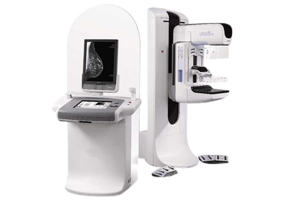
It is a diagnostic method used to detect breast cancer and reduce breast cancer-related deaths by examining breast tissue with low-dose X-rays.

More information
4D Mammography
Mammography

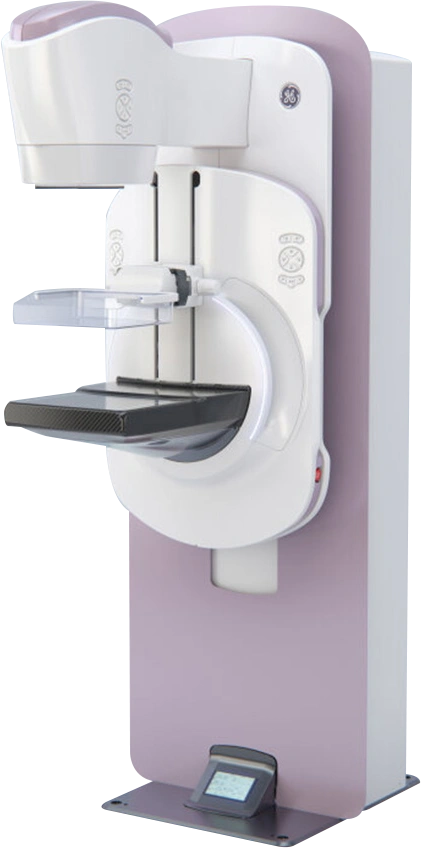
Mammography is the most effective imaging method for early diagnosis of breast cancer, which ranks as the second leading cause of death among women.

More information
Mammography
ECG

It is a quick and painless test commonly used for the detection of certain heart diseases.

More information
ECG
Endobronchial Ultrasound (EBUS)

Endobronchial Ultrasound (EBUS), performed at only a few centers in Turkey, is a diagnostic method with low complication risks. What sets it apart is its ability to simultaneously diagnose and stage lung cancer in a single session, minimizing time loss for the patient.

More information
Endobronchial Ultrasound (EBUS)
Healthy Teeth with Three-Dimensional Oral Scanning Technology

Technological advancements are providing significant opportunities in dental care, as in other fields. Dr. Arzu Tekkeli, Clinical Coordinator of the Oral and Dental Health Department at Anadolu Sağlık Merkezi, explains that "three-dimensional oral scanning allows for the internal structure of the mouth to be scanned and images to be transferred to a computer, enabling the production and placement of a new tooth with almost zero error on the same day. Three-dimensional oral scanning, achieved through the digital integration of three different devices, quickly resolves issues such as missing teeth, tooth alignment, broken teeth, fillings, or crowns in just one day. This means patients do not have to make multiple visits to the clinic for treatment. The same method also provides quick and long-lasting results for many other dental problems." Dr. Arzu Tekkeli provided important insights on the topic.

More information
Healthy Teeth with Three-Dimensional Oral Scanning Technology
MR Linac

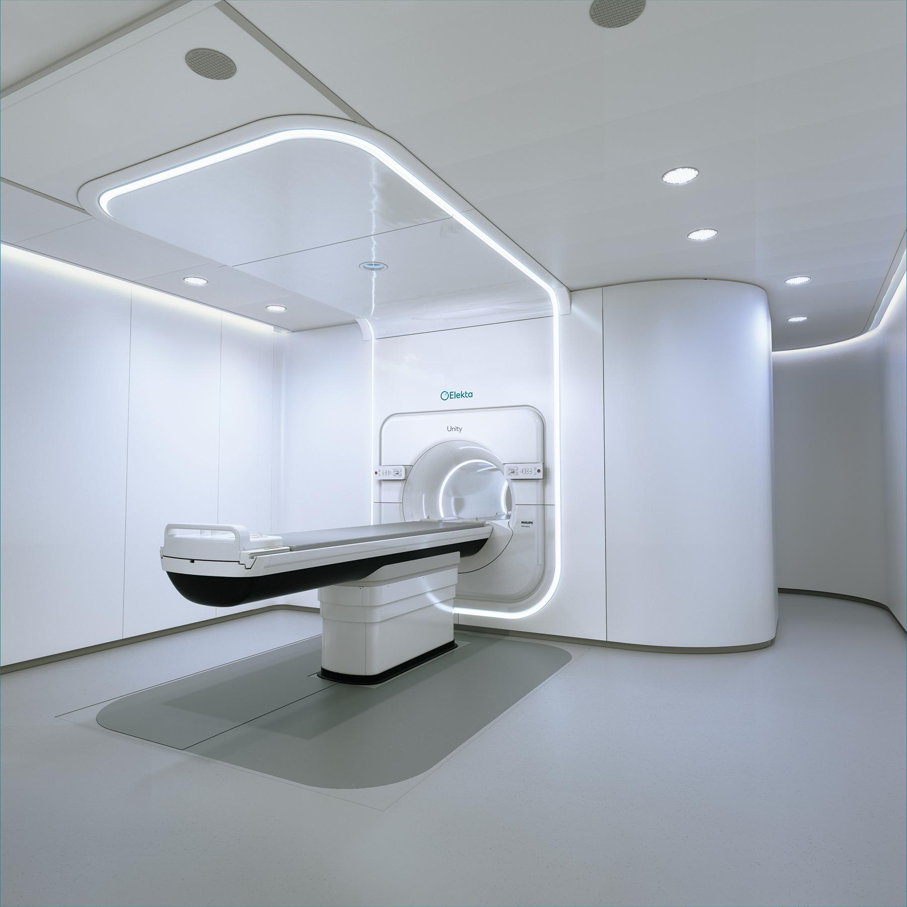
Significant advancements are being made in cancer treatments today. One of the areas experiencing the most important developments is Radiation Oncology.

More information
MR Linac
StemWave

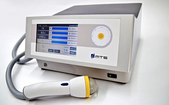
Tissue Regeneration Technology (TRT) is based on the principle of using high-energy sound waves (mechanoacoustic) generated by a special liquid and electrical arc system, which are directly delivered to the damaged, painful area via a transducer (mechanotransduction). The successive high-energy sound waves stimulate the healing process at the cellular, tissue, and systemic levels through natural effects in the treated area.

More information
StemWave
Sialendoscopy Method (Salivary Gland Endoscopy)

Sialendoscopy is an endoscopic procedure that involves visualizing the salivary gland duct and diagnosing and treating salivary gland diseases. The method first diagnoses the pathology within the duct, and then endoscopic treatment is performed either in the same session or in a subsequent session.

More information
Sialendoscopy Method (Salivary Gland Endoscopy)
Computed Tomography

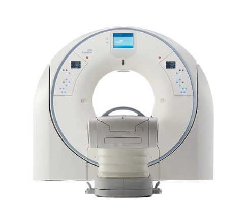
Fast and clear imaging: We provide high-resolution images that accelerate disease diagnosis with artificial intelligence and deep learning applications.

More information
Computed Tomography

Ear, Nose, Throat Diseases Department

Department Doctors

Gebze

Ear, Nose, Throat Diseases Department

Prof. Bülent Evren Erkul

Gebze

Ear, Nose, Throat Diseases Department

Prof. Ziya Saltürk

Gebze

Ear, Nose, Throat Diseases Department

MD. Surgeon Ahmet Hamdi Önay

Gebze

Language and Speech Therapy Department

Therapist Kayra Göktaş

Show All
Gebze

Ear, Nose, Throat Diseases Department

Prof. Bülent Evren Erkul

Gebze

Ear, Nose, Throat Diseases Department

Prof. Ziya Saltürk

Gebze

Ear, Nose, Throat Diseases Department

MD. Surgeon Ahmet Hamdi Önay

Gebze

Language and Speech Therapy Department

Therapist Kayra Göktaş

Show All