Ultrasonography

Ultrasound, also known as ultrasonography, is a medical imaging technique that uses high-frequency sound waves to produce images. With advancements in technology, ultrasound devices have greatly improved, allowing for 2D, 3D, and 4D imaging, which enables more accurate diagnoses by specialists. Ultrasound is commonly used to evaluate various organs, detect tumor lesions, and monitor pregnancy. By gaining detailed information about ultrasound terms and methods, you can better understand the process. 
What is Ultrasound and Detailed Ultrasound? 

The question of what ultrasound is often arises for those who need to undergo an ultrasound or their relatives. Ultrasound is a procedure that allows for the evaluation of organs inside the body, detection of changes, developments, and problems. This method enables easy detection of issues by allowing for a thorough observation of the current situation.  

Ultrasound is also used to monitor the development of the fetus during pregnancy, observe issues in soft tissues, and diagnose problems in internal organs. Ultrasound is performed in two different ways: standard 2D ultrasound and detailed ultrasound. The most frequently asked questions about ultrasound types include what is standard ultrasound, what is detailed ultrasound, and what is detailed ultrasound. Standard ultrasound is a 2D imaging procedure performed at any time during pregnancy. Since very clear images cannot be obtained in the early months, specialists typically perform 2D ultrasound from the 16th week onward.  

Detailed ultrasound is also frequently used during pregnancy. Also known as detailed ultrasound, this method provides detailed information about the baby's health and organs, allowing for a thorough monitoring of the baby's development. 

Additionally, this method is used to examine cancerous tissues in organs, assess tissues, and detect the presence of any tumor masses that might exist.  

What Are the Types of Ultrasound Devices? 

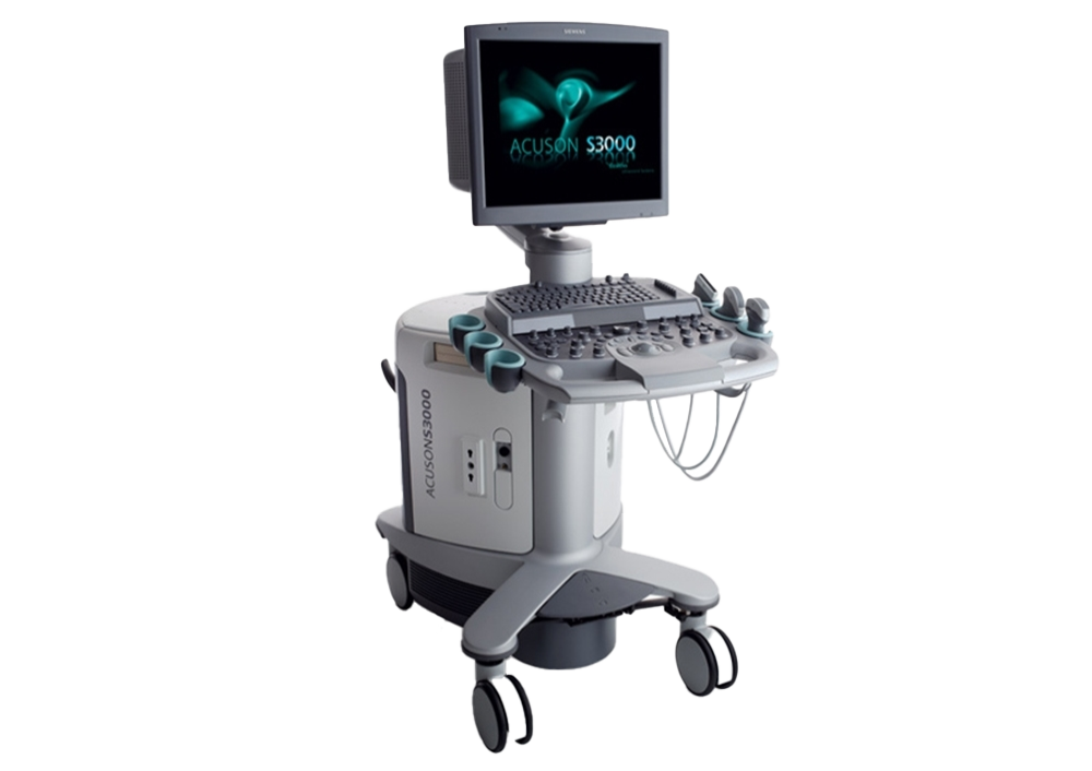
The medical device that allows for the examination of changes in the human body that are not visible to the eye is called ultrasonography. Commonly referred to as ultrasound, this device uses sound waves at frequencies inaudible to the human ear to move through tissues. The real-time images obtained with these sound waves can be monitored via a connected monitor. The ultrasound device consists of two separate parts. The first part is the probe that comes into contact with the body and is manually controlled. The other part is the image processing unit that makes the sound waves sent by the probe visible. Ultrasound devices are classified into different types based on the quality and detail of the images they provide. 

  • 2D Ultrasound Device 

2D ultrasound devices are primarily used for routine examinations. 2D ultrasound machines are used for general assessment and monitoring purposes. In pregnancy monitoring, 2D ultrasound devices are commonly used to easily visualize the baby's heartbeat, the position of the placenta, and the baby's umbilical cord. The images obtained from these devices are black and white. The baby's length, weight, and physical development can also be monitored with 2D ultrasound devices. Additionally, 2D ultrasound applications are the most cost-effective option in terms of ultrasound prices. 

  • 3D Ultrasound Device 

3D ultrasound devices allow for clearer results, especially in pregnancy monitoring. The 3D ultrasound application enables detailed tracking of the baby's development at various weeks. Additionally, 3D ultrasound provides a close-up, larger, and colored image of the baby. Typically used after the 3rd month, 3D ultrasound makes it easier to understand the baby's weight, length, and volume. Since 3D ultrasound provides a detailed image of whether the baby is male or female, it facilitates gender determination. 

  • 4D Ultrasound Device 

4D ultrasound devices are equipped with technology that allows for three-dimensional and moving images. Especially used during pregnancy, 4D ultrasound machines enable the monitoring of the baby's head, foot, and hand movements like a film from the womb. With 4D devices, in addition to determining the sex, spine development, hands, nose, skull bones, and internal organs can be viewed in great detail. This allows for planning post-birth interventions or treatments in detail if there are concerns about specific areas. Moreover, detailed ultrasound with these devices can detect certain problems in internal organs that might be missed with other types of ultrasound devices. 

Medical Technologies

All Technologies
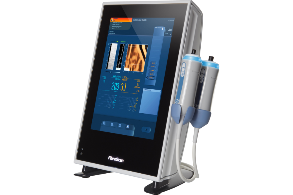
Fibroscan

The gold standard in the assessment of liver diseases is liver biopsy. However, thanks to the newly-devised Fibroscan technology, ultrasound waves are now used instead of biopsies in many liver diseases.

More information
Fibroscan
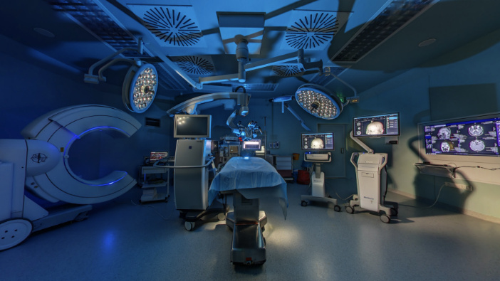
Hybrid Operating Room

"Hybrid operating room" refers to a new concept of operating rooms where advanced medical imaging systems and medical devices can be used simultaneously.

More information
Hybrid Operating Room
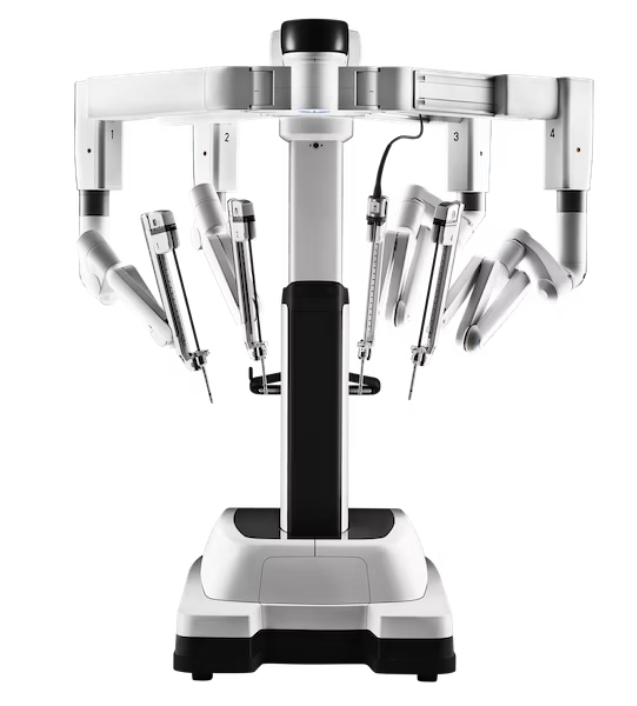
Robotic Surgery

Robotic surgery or robot-assisted surgery, "da Vinci" system of small holes can be defined as the realization of some operations.

More information
Robotic Surgery
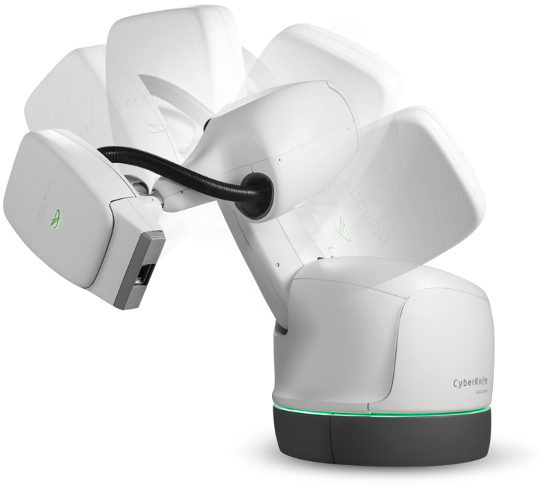
CyberKnife M6

Eliminate tumor surgically lifting up a certain limit, it is a system that prevents the harm of radiation to normal tissues.

More information
CyberKnife M6
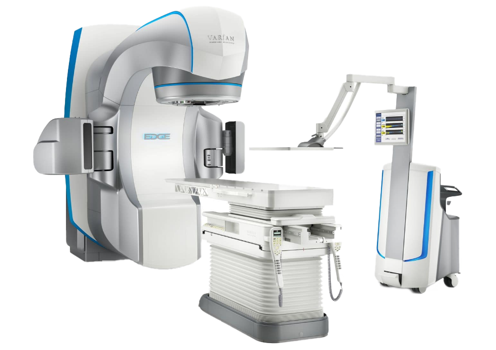
Varian Edge

The Varian Edge, first used in Turkey at Anadolu Sağlık Merkezi, is one of the advanced radiotherapy devices that combines the most advanced technologies in cancer radiotherapy.

More information
Varian Edge
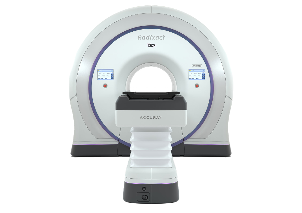
Radixact

The Radixact device, produced by Accuray, is the newest model in the "tomotherapy" device family.

More information
Radixact
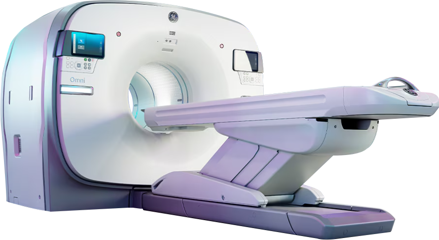
PET-CT

PET/CT, PET (Positron Emission Tomography) and CT (Computed Tomography) techniques combining in the same device is an imaging method that provides more information than they give them separately.

More information
PET-CT
Intraoperative Radiotherapy

In the treatment of breast cancer, radiotherapy today focuses on delivering lower intensity, lower doses, to fewer areas, and for shorter durations.

More information
Intraoperative Radiotherapy
MR-TRUS Fusion

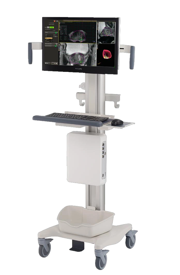
Tumor targeted MR-TRUS Fusion Biopsy is a method, which detects foci suspicious of prostate cancer, and ensures taking biopsy from these foci with millimetric sensitivity by means of an advanced technology device and software.

More information
MR-TRUS Fusion
Radiology Equipment

Radiology Equipment

More information
Radiology Equipment
Ultrasonography

Ultrasound, or more formally, ultrasonography, is a medical imaging technique that uses high-frequency sound waves to produce images.

More information
Ultrasonography
4D Mammography

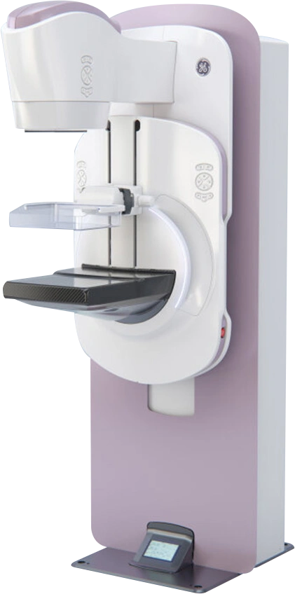
It is a diagnostic method used to detect breast cancer and reduce breast cancer-related deaths by examining breast tissue with low-dose X-rays.

More information
4D Mammography
Mammography

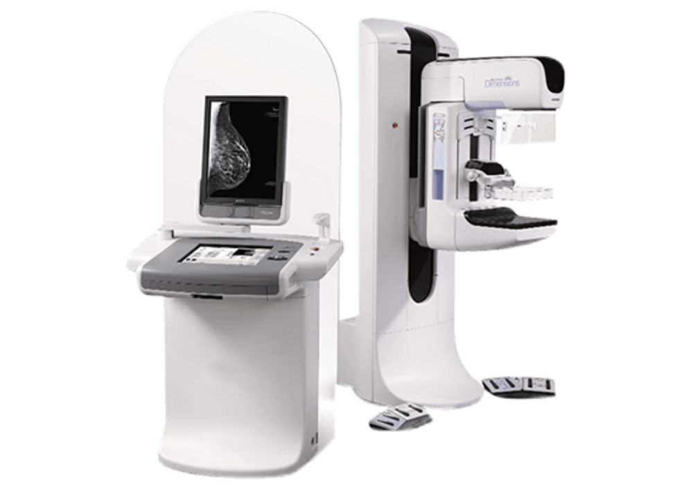
Mammography is the most effective imaging method for early diagnosis of breast cancer, which ranks as the second leading cause of death among women.

More information
Mammography
ECG

It is a quick and painless test commonly used for the detection of certain heart diseases.

More information
ECG
Endobronchial Ultrasound (EBUS)

Endobronchial Ultrasound (EBUS), performed at only a few centers in Turkey, is a diagnostic method with low complication risks. What sets it apart is its ability to simultaneously diagnose and stage lung cancer in a single session, minimizing time loss for the patient.

More information
Endobronchial Ultrasound (EBUS)
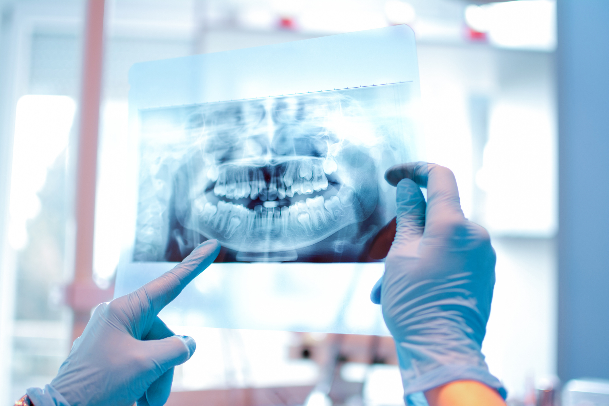
Healthy Teeth with Three-Dimensional Oral Scanning Technology

Technological advancements are providing significant opportunities in dental care, as in other fields. Dr. Arzu Tekkeli, Clinical Coordinator of the Oral and Dental Health Department at Anadolu Sağlık Merkezi, explains that "three-dimensional oral scanning allows for the internal structure of the mouth to be scanned and images to be transferred to a computer, enabling the production and placement of a new tooth with almost zero error on the same day. Three-dimensional oral scanning, achieved through the digital integration of three different devices, quickly resolves issues such as missing teeth, tooth alignment, broken teeth, fillings, or crowns in just one day. This means patients do not have to make multiple visits to the clinic for treatment. The same method also provides quick and long-lasting results for many other dental problems." Dr. Arzu Tekkeli provided important insights on the topic.

More information
Healthy Teeth with Three-Dimensional Oral Scanning Technology
MR Linac

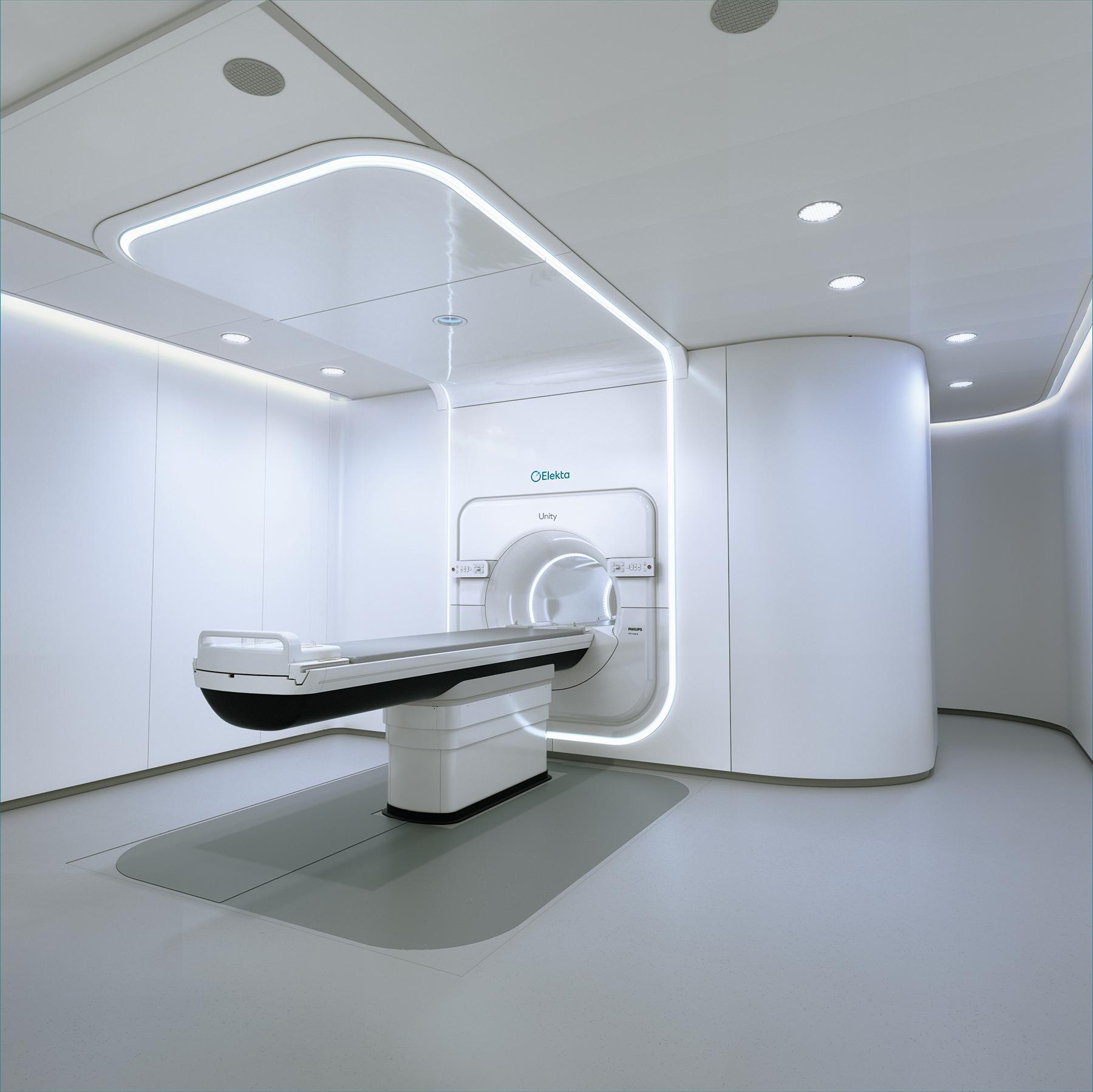
Significant advancements are being made in cancer treatments today. One of the areas experiencing the most important developments is Radiation Oncology.

More information
MR Linac
StemWave

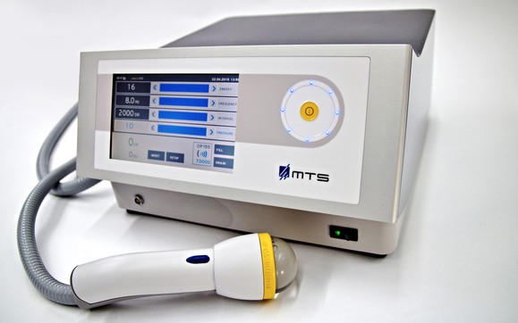
Tissue Regeneration Technology (TRT) is based on the principle of using high-energy sound waves (mechanoacoustic) generated by a special liquid and electrical arc system, which are directly delivered to the damaged, painful area via a transducer (mechanotransduction). The successive high-energy sound waves stimulate the healing process at the cellular, tissue, and systemic levels through natural effects in the treated area.

More information
StemWave
Sialendoscopy Method (Salivary Gland Endoscopy)

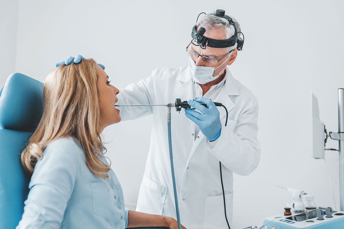
Sialendoscopy is an endoscopic procedure that involves visualizing the salivary gland duct and diagnosing and treating salivary gland diseases. The method first diagnoses the pathology within the duct, and then endoscopic treatment is performed either in the same session or in a subsequent session.

More information
Sialendoscopy Method (Salivary Gland Endoscopy)
Computed Tomography

Fast and clear imaging: We provide high-resolution images that accelerate disease diagnosis with artificial intelligence and deep learning applications.

More information
Computed Tomography

Breast Center

Department Doctors

Gebze

Breast Center

Prof. Ali Uğur Emre

Gebze

Breast Center

Assoc. Prof. Ayhan Erdemir

Breast Center

Assoc. Prof. Özgür Sarıca

Gebze

Breast Center

MD. Surgeon Kemal Raşa

Gebze

Medical Oncology

Prof. Bülent Karagöz

Gebze

Medical Oncology

Prof. Necdet Üskent

Gebze

Medical Oncology

Prof. Şeref Kömürcü

Gebze

Medical Oncology

Prof. Yeşim Yıldırım

Gebze

Medical Oncology

Assoc. Prof. Eda Tanrıkulu Şimşek

Gebze

Medical Oncology

MD. Sinan Karaaslan

Gebze

Radiation Oncology Department

Prof. Hale Başak Çağlar

Gebze

Radiation Oncology Department

MD. Mehmet Doğu Canoğlu

Gebze

Radiation Oncology Department

MD. Rashad Rzazade

Gebze

Radiology Department

MD. Adnan Aras

Gebze

Radiology Department

MD. Ahmet Murat Dökdök

Gebze

Radiology Department

MD. Emine Okur

Gebze

Radiology Department

MD. Kutlay Karaman

Gebze

Radiology Department

MD. Oktay Karadeniz

Ataşehir

Radiology Department

MD. Yeter Tatlıadım

Gebze

Thoracic Surgery Department

Prof. Altan Kır

Gebze

Urology Department

Prof. İlker Tinay

Show All
Gebze

Breast Center

Prof. Ali Uğur Emre

Gebze

Breast Center

Assoc. Prof. Ayhan Erdemir

Breast Center

Assoc. Prof. Özgür Sarıca

Gebze

Breast Center

MD. Surgeon Kemal Raşa

Gebze

Medical Oncology

Prof. Bülent Karagöz

Gebze

Medical Oncology

Prof. Necdet Üskent

Gebze

Medical Oncology

Prof. Şeref Kömürcü

Gebze

Medical Oncology

Prof. Yeşim Yıldırım

Gebze

Medical Oncology

Assoc. Prof. Eda Tanrıkulu Şimşek

Gebze

Medical Oncology

MD. Sinan Karaaslan

Gebze

Radiation Oncology Department

Prof. Hale Başak Çağlar

Gebze

Radiation Oncology Department

MD. Mehmet Doğu Canoğlu

Gebze

Radiation Oncology Department

MD. Rashad Rzazade

Gebze

Radiology Department

MD. Adnan Aras

Gebze

Radiology Department

MD. Ahmet Murat Dökdök

Gebze

Radiology Department

MD. Emine Okur

Gebze

Radiology Department

MD. Kutlay Karaman

Gebze

Radiology Department

MD. Oktay Karadeniz

Ataşehir

Radiology Department

MD. Yeter Tatlıadım

Gebze

Thoracic Surgery Department

Prof. Altan Kır

Gebze

Urology Department

Prof. İlker Tinay

Show All