Hybrid Operating Room
What is a Hybrid Operating Room?
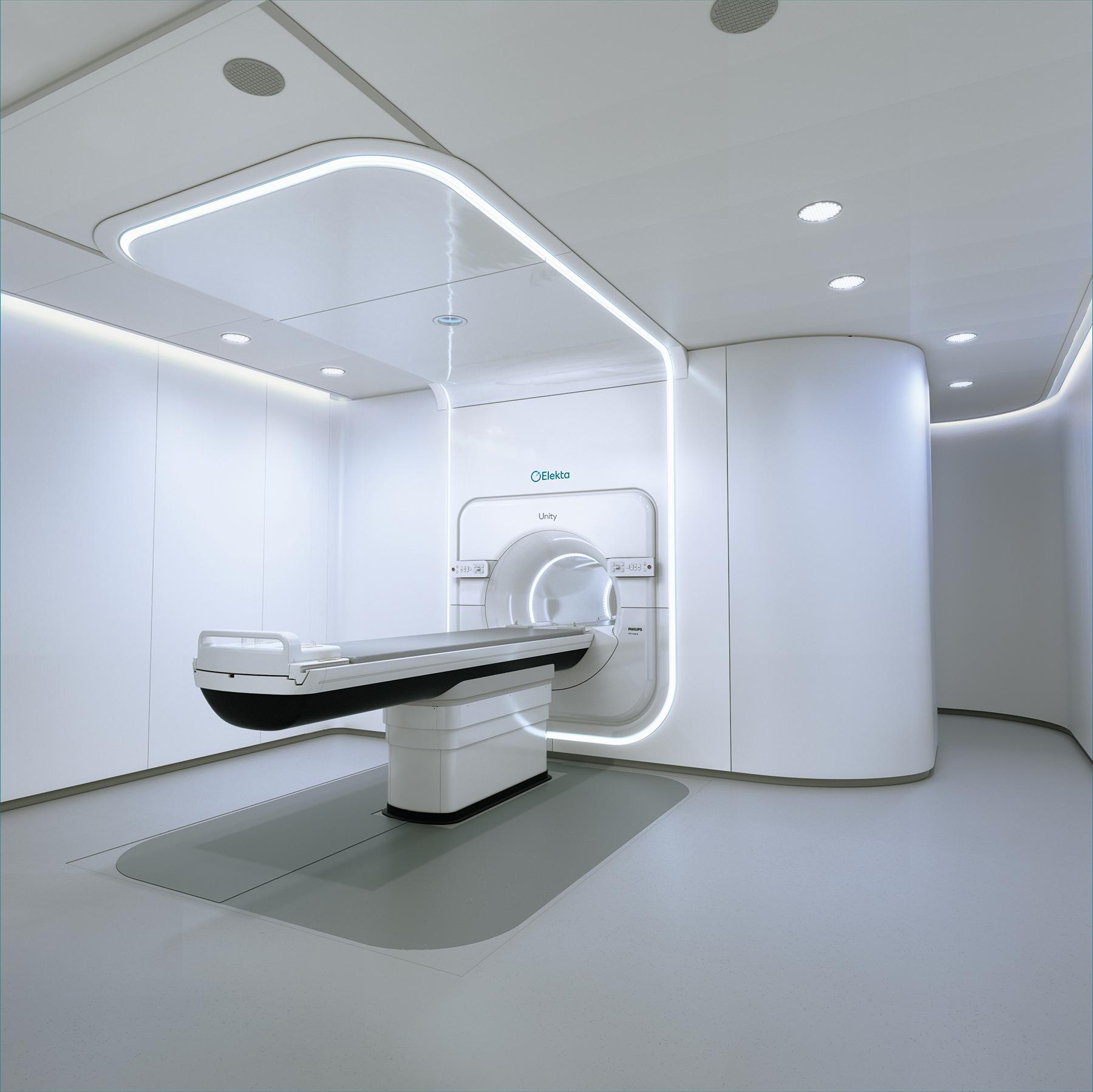
The term "hybrid operating room" refers to a new concept of operating room where advanced medical imaging systems and medical devices can be used simultaneously. It started with the use of mobile X-ray devices, known as C-arm fluoroscopy, in operating rooms in the 1970s. Today, the process continues with the introduction of mobile computed tomography (CT) and magnetic resonance (MR) imaging devices, and has reached a fully hybrid concept with the addition of advanced technologies such as neuronavigation (surgical target determination) and neuromonitoring devices (monitoring the functions of the brain and nerves during surgery). Recently, the integration of special fluorescent-filtered surgical microscopes with navigation systems into the medical equipment of the operating room has made it easier to distinguish tumors from normal nerve tissues, and microscopes have also started to be included in the "hybrid" concept.
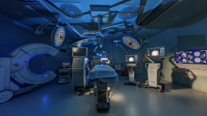
The development of hybrid operating rooms has coincided with the development of minimally invasive surgical techniques in our country and around the world. Therefore, the most difficult and problematic complex surgeries can be performed more safely. This brings advantages such as less surgical trauma, smaller incisions, shorter operation times, less blood loss, fewer complications, shorter hospital stays, and reduced costs for both the patient and the surgeon.
In neurosurgery, spine, and spinal cord surgeries, "Hybrid Operating Rooms" are used for many disease groups and trauma surgeries. Some of these include neurovascular surgery for brain or spinal vascular diseases such as aneurysms and arteriovenous malformations (AVM), brain tumors, functional stereotactic surgery, and spinal implantation surgery.
Technologies Used in the Hybrid Operating Room:
The health technologies used in our Hybrid Operating Rooms include:
- O-ARM CT
- Neuro-Navigation System
- Next-Generation Fluorescent-Filtered Microscope
- Intraoperative Neuromonitoring
O-ARM CT
O-ARM CT (O-ARM TOMOGRAPHY)
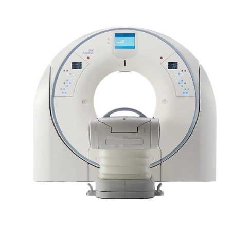
With the new generation imaging system, the mobile tomography device, it is possible to perform a computed tomography scan during surgery. This technology allows for 2D or 3D imaging of both the brain and spine (in spine surgery). Imaging can be done in 13 seconds in a 360° and 3D manner, providing information about the relevant area of the patient on the operating table. Thanks to the system's robotic positioning feature, it can be quickly brought to the patient table in the operating room, and tomography images can be obtained. Additionally, since it captures images in a single rotation, the same image quality can be observed during the surgery with one-third of the radiation dose compared to other standard tomography devices.
It is frequently used in surgeries for placing screws (platin placement) in spinal deformities due to aging, spinal canal compressions, spinal tumors, scoliosis in childhood and adolescence, certain developmental diseases of the spine, and fractures and dislocations caused by trauma.
The placement of screws in the spine must be determined with 1-2 mm accuracy, depending on the anatomical region. Until recently, spinal screw surgeries were performed using C-arm and 2D imaging, known as fluoroscopy. In these surgeries, due to the possibility of screws being misplaced, patients might need to undergo surgery again, increasing the risk of infection and prolonging the hospital stay. However, with the O-Arm CT technology that allows 3D tomography imaging, the success rate of screw surgeries has increased, and the margin of error has been eliminated. Additionally, the neuro-navigation system, which works in harmony with the O-Arm CT device and can display all targets with high precision, is used simultaneously, further enhancing the success rate of the surgery. Therefore, surgery can be performed safely, minimizing the risk of spinal cord and nerve root injuries to nearly zero with 1-2 mm accuracy.
Thanks to the O-Arm CT system, unwanted situations that require the patient to undergo another surgery, such as misplaced screws in spinal surgeries or bleeding in the surgical area in brain surgeries, are minimized by taking a tomography scan under sterile conditions at the final stage of the surgery.
Advantages of Spine Screw Surgeries Performed with the O-Arm Device;
- Provides critical information to the surgeon at every stage, reducing the risk of reoperation.
- Patient receives less radiation.
- Allows for quicker recovery with a smaller surgical incision and reduces bleeding.
- This system minimizes the major risks associated with complex surgeries.
- Reduces the risk of infection. The risk of paralysis due to screws is minimized.
Neuro-Navigation System
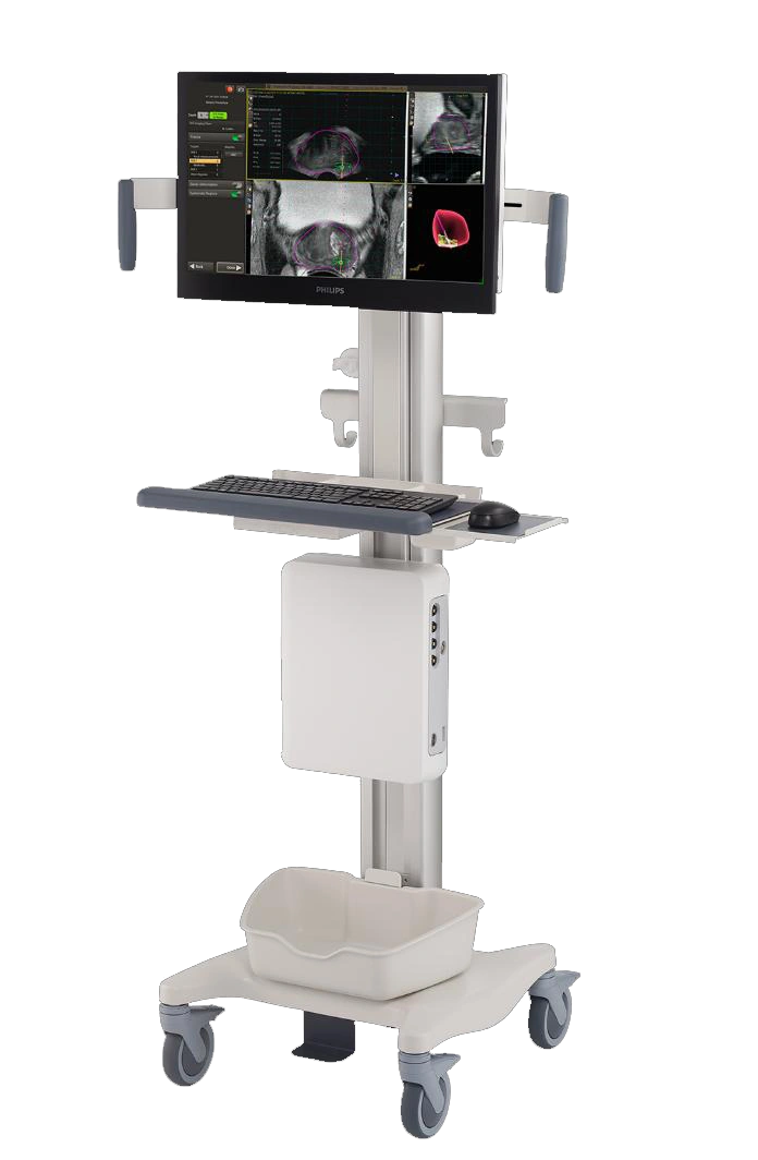
The new generation neuro-navigation is a "navigation or guidance" system. It calculates coordinates using a system similar to GPS technology for a target in the brain or spine surgery and allows the surgeon to see the 3D scanned images. With "neuro-navigation," which is a high-level design of computer technology, the surgeon can approach the targeted lesion in brain surgery with high accuracy (less than 1 mm) and minimize damage to healthy tissue during surgery. In this method, the patient's MR and/or CT is taken before the surgery and transferred to the navigation device. Thus, during the surgery, various risk areas in the patient's brain and spine can be seen in real-time navigation, and planning can be done accordingly. The neuro-navigation system calculates the coordinates of the surgical area, reaching the target with minimal deviation. This increases surgical success and minimizes the risks of death and disability.
This method can be used alone or in combination with other technologies. In cases of small and deep-seated tumors and similar conditions, stereotactic biopsy can be performed using a small entry hole, i.e., a tissue sample can be taken by reaching the area millimetrically with a fine needle. Based on the pathological examination of this tissue sample, the treatment method for the patient is determined.
Advantages of the Neuro-Navigation System;
- Progresses flawlessly over sensitive anatomy, avoiding critical structures.
- Surgery is performed within more minimally invasive limits (surgery time is shortened with a small surgical incision).
- Provides a safer surgery option by not disrupting the patient's healthy anatomy.
Next-Generation Fluorescent-Filtered Microscope
The treatment of brain and spinal cord tumors is microsurgery. The complete removal of tumors (increasing the extent of resection) is the most important factor affecting patients' prognosis and survival. The biggest challenge in tumor surgery is distinguishing the tumor tissue from normal tissue. Achieving this with a standard normal surgical microscope can sometimes be problematic. However, with next-generation microscopes, using special light filters, tumor tissue stained with fluorescence can be easily distinguished from normal nerve tissue. Therefore, with the special surgical microscope combined with neuro-navigation, the maximum removal (resection) of the tumor with minimal error can be achieved with a smaller incision. It is a system found only in certain hospitals in Turkey. In addition to tumor surgeries, during surgeries for brain vascular diseases, the microscope filter can be adjusted to show the brain's vessels by injecting a special substance into the patient during the surgery, allowing brain vascular angiography. In this way, the vascular network of the brain can be seen before the surgery is completed. This increases the chance of safely surgically treating brain vascular disorders.
Fluorescent-Guided Tumor Surgery is successfully applied in our hospital's Brain and Spinal Cord Tumor Center in hybrid operating room conditions with intraoperative imaging, neuro-navigation, and neuromonitoring technologies.
Intraoperative Neuromonitoring
In order to minimize nerve damage (risk of paralysis) that may develop due to brain tumor and spinal surgeries, the system continuously monitors the brain, spinal cord, nerve roots, and reflex pathways on the monitor by applying electrical stimuli through electrodes placed on the patient's scalp and muscles during the surgery. When a change is detected in the values monitored by the neurologist throughout the surgery, the surgeon is warned. Thus, irreversible damage (such as paralysis) in the patient is prevented, and the quality of life is maintained. The patient can quickly return to home and work life.


