Robotic Surgery
What is Robotic Surgery?
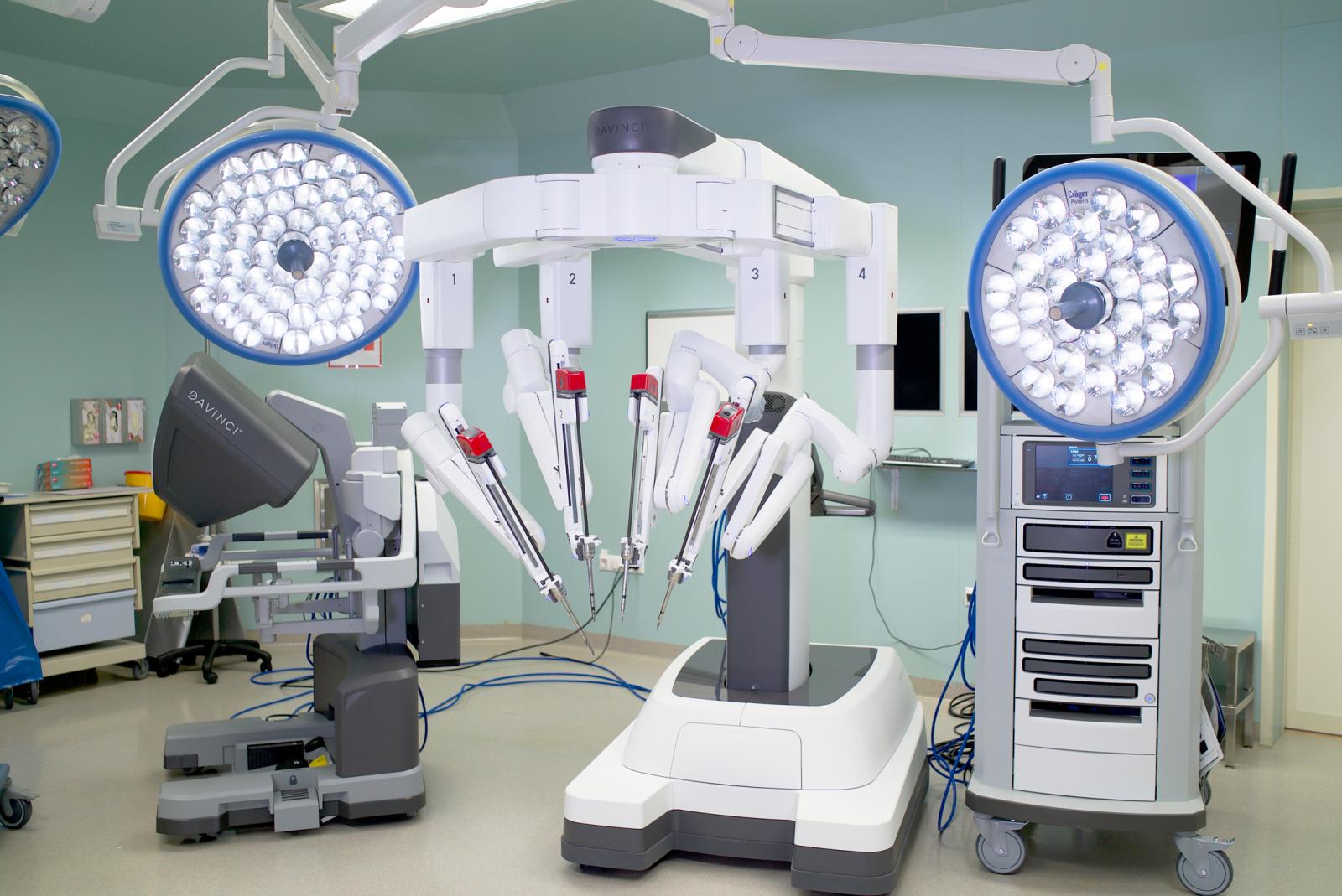
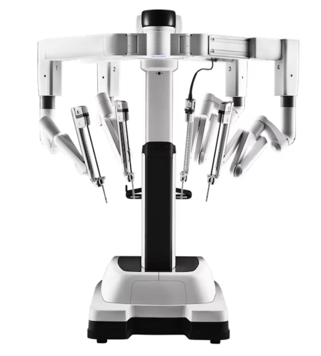
Robotic surgery or robot-assisted surgery is defined as performing certain surgeries through small incisions using the "da Vinci" system. As the next step in minimally invasive surgery following traditional surgeries and laparoscopic surgery, it offers numerous conveniences for both patients and surgeons.
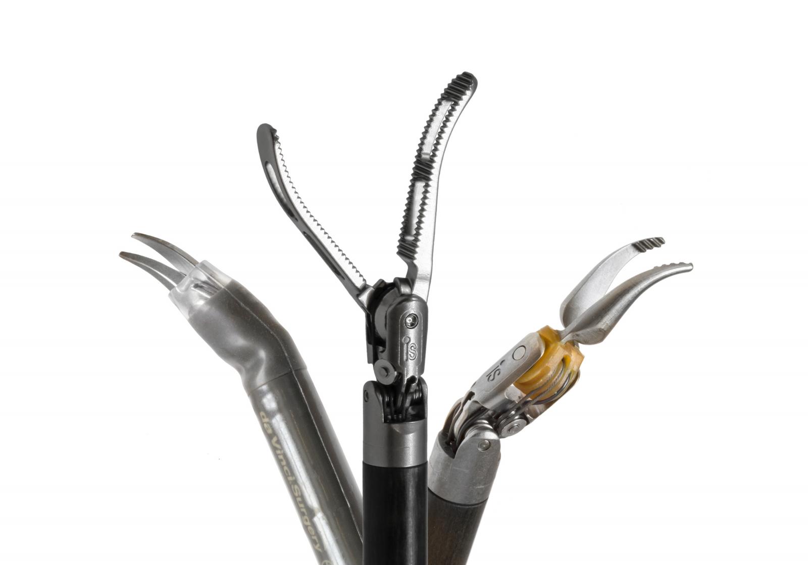
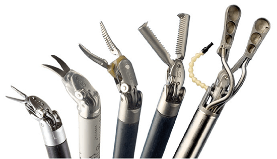
Instruments modeled on the human wrist, intuitive motion control, and high-definition three-dimensional imaging allow the surgeon to overcome limitations in traditional open or closed surgical technologies, enabling more complex interventions through minimally invasive surgery. Thanks to this method, the surgeon seated at the console can view a clear, three-dimensional image of the surgical area.
What is minimally invasive surgery?
In minimally invasive surgery, the goal is to access the affected area with the smallest incision possible, without harming bones and tissues, and to perform the necessary intervention. Such surgeries mean shorter hospital stays and faster recovery for patients. Other benefits may include:
- Less blood loss
- Less scarring
- Reduced risk of infection
- Accelerated recovery process
What are the advantages of the robotic surgery method?
Compared to traditional open surgery and laparoscopic closed surgery approaches, robotic surgery has several advantages. For suitable patients and cases, robotic surgery is preferred due to the following features:
- Surgeons can see the surgical area more clearly and with greater precision thanks to the camera on the robotic arm, which increases the success of the surgery.
- The use of robotic arms allows surgical procedures to be performed more easily and effectively.
- The robotic system enables simultaneous operation of multiple arms, allowing for the execution of techniques that are typically complex or impossible.
- Robotic surgery, which can be performed through a small incision, provides the advantages of minimally invasive surgery. This method reduces the likelihood of complications associated with the surgical area. It results in less pain and blood loss after surgery, speeds up recovery, and leaves a smaller scar.
- The hospital stay after robotic surgery is significantly reduced, allowing the patient to be discharged early and start post-surgical treatments, such as medication for cancer surgeries, sooner.
- Important tissues in the surgical area, such as nerves and blood vessels, can be more easily identified thanks to the high-definition camera on the robotic arm, allowing the surgeon to examine the area comprehensively and in depth. This minimizes the risk of damage to surrounding tissues during the surgery, reducing the chance of postoperative complications.
- Thanks to high-resolution imaging, repairs on small tissues and vessels that cannot be easily seen by the human eye can be performed with ease.
- The ergonomic design of the robotic system enhances the surgeon's comfort during lengthy surgical procedures, positively impacting surgical success.
- In robotic surgeries, geometrically precise and accurate repairs result in more satisfying outcomes, both functionally and cosmetically.
- Robotic arms can be sterilized more effectively than human hands and carry no biological risks, ensuring a more sterile and safe surgical field.
Robotic Surgery in General Surgery
One of the fields where robotic surgery systems are widely used is general surgery. As in other procedures, robotic surgery systems provide additional benefits for both doctors and patients in selected general surgery operations. Similar to other surgeries performed with da Vinci, robotic surgery in general surgery offers advantages for patients, such as fewer incisions and therefore less pain, bleeding, and shorter recovery times. The primary benefit, however, is the ability to see and access deep tissues that are difficult to reach and view with traditional methods, allowing for more detailed and flawless procedures.
Among the most successful surgeries performed with robotic systems are colon and rectum (large intestine) surgeries.
General surgical procedures that can be performed with the da Vinci robot include:
- Colorectal diseases, especially cancers,
- Stomach and esophagus diseases,
- Pancreatic diseases,
- Spleen diseases,
- Liver and biliary tract diseases,
- Some endocrine organ diseases, especially of the adrenal glands,
- Obesity surgery
Robotic Surgery in Urological Diseases
Urology is one of the fields where the da Vinci robotic surgery system is most commonly used. Due to the advantages it provides to both the patient and the doctor, the frequency of robotic-assisted surgery applications is steadily increasing. The most common application of this system worldwide is in the surgical treatment of prostate cancer, the most common cancer in men, through radical prostatectomy (removal of the prostate), and it has been successfully implemented.

Other applications of robotic-assisted surgery in urological oncology:
- Radical nephrectomy (removal of the entire kidney) and partial nephrectomy (removal of the tumor portion of the kidney) for patients diagnosed with kidney cancer,
- Radical cystectomy and creation of a neobladder (artificial bladder) for patients diagnosed with bladder cancer,
- Radical nephroureterectomy (removal of the kidney and ureter together) for upper urinary system cancers,
- Removal of retroperitoneal masses in patients diagnosed with testicular cancer,
Other common applications in urology:
- Pyeloplasty (correction of uretero-pelvic strictures)
- Removal of bladder diverticula (bladder diverticulectomy),
- Robotic surgery for pyeloplasty-ureteropelvic junction strictures,
- Surgical treatments for bladder prolapse in women,
Robotic-assisted surgeries can be performed with great precision at every stage, thanks to features such as the 3D imaging transferred to the console and the robot’s ability to move in multiple directions.
Robotic Surgery in Gynecological Diseases
In gynecological surgery, a significant number of complex operations, including cancer surgeries, hysterectomy (removal of the uterus), removal of large fibroids, correction and reconstruction of genital prolapse, and tubal reanastomosis (reconnecting fallopian tubes after ligation), can be performed with robotic surgery systems. Robotic technology provides the ability to perform suturing comfortably inside the abdomen, even enabling the tools to rotate beyond what the wrist can manage. The 10x magnified detailed imaging greatly assists the surgeon.
The most common surgeries in the field of gynecology include cancer surgeries, hysterectomy (removal of the uterus), myomectomy (removal of fibroids), sacrocolpopexy (lifting a prolapsed vagina), and tubal reanastomosis surgery (reconnecting fallopian tubes after ligation).
Robotic surgeries that can be performed in gynecology with the da Vinci robot:
- Robotic surgery in uterine and cervical cancer
- Robotic surgery in fibroid surgeries
- Other gynecological surgical procedures
Robotic Surgery in Thoracic Surgery
Thoracic Surgery primarily deals with the surgical diseases of the lungs, but it also involves surgeries of the main airways, interstitial space between the lungs called the mediastinum, pleura (lining of the lungs and chest cavity), esophageal surgeries, chest wall deformities and tumors, and surgeries of the diaphragm, which separates the chest cavity from the abdominal cavity.

As in many other surgical branches, both open and closed surgeries are performed in thoracic surgery. Open surgery is a classical procedure that involves making an incision in the chest, the length of which varies depending on the size of the surgery. Closed surgeries, also known as "minimally invasive surgery," are performed with minimal harm to the patient. However, not all patients are suitable for closed surgical interventions. The decision to perform a closed surgery depends on the organ affected, the type and size of the tumor, and its relation to surrounding tissues or organs. Nowadays, there are two types of closed surgeries in thoracic surgery. The first is the video-assisted thoracoscopic surgery (VATS) method, which started in the 1990s. It is performed through one or more small incisions made in the chest cavity. In this procedure, the surgeon looks at a monitor and performs the surgery using surgical instruments inserted through these incisions. The image is two-dimensional, and the maneuverability of the instruments is limited.

Video-assisted thoracoscopic surgery (VATS)
After the 2000s, with the advancement of robot technology, robots started to be used in surgeries. The Da Vinci robotic systems are now widely used in surgery.
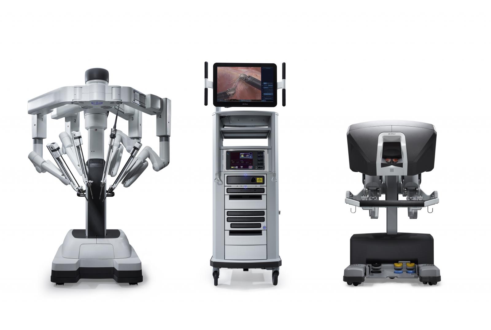
These robots are most commonly used in urology, thoracic, and abdominal surgeries. In Robotic-Assisted Thoracoscopic Surgery (RATS), four 8mm incisions and one 15mm incision are made in the patient's chest cavity. The first four incisions are used for the robot's arms, and the largest incision is for the assistant. After positioning the robot near the patient, the surgical instruments are attached to three of its arms, and a camera is mounted on the fourth arm.

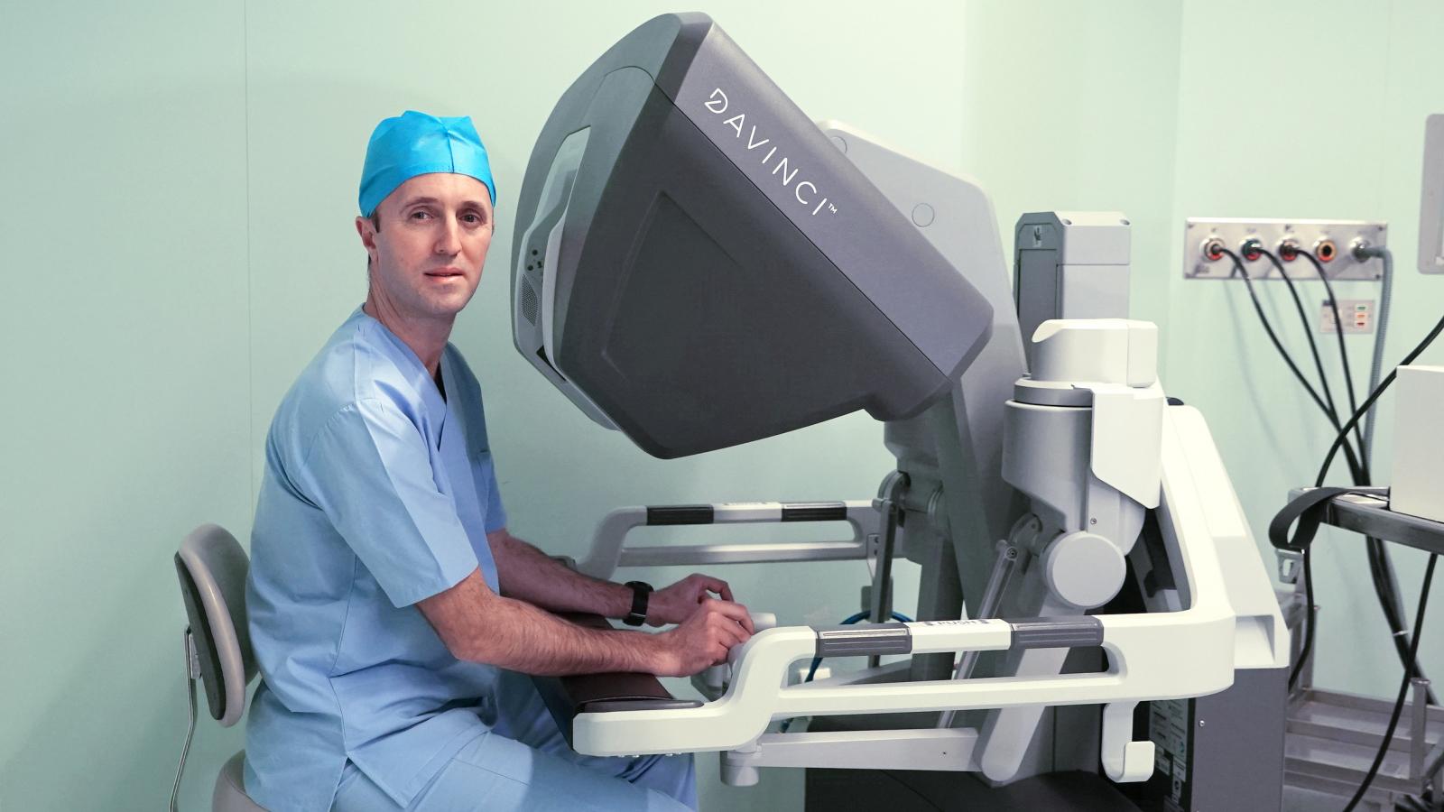
At this stage, the surgeon leaves the patient's bedside and sits at a console. From this console, the surgeon can view the patient's interior in 3D and perform the procedure by controlling joystick-like instruments with their fingers.
Among the most common surgeries in the field of gynecology are cancer surgeries, hysterectomy (removal of the uterus), myomectomy (removal of fibroids), sacrocolpopexy (lifting of the prolapsed vagina), and tubal reanastomosis surgery (restoring the function of the tubes after tubal ligation).
Surgeries in Gynecology that can be performed with the da Vinci robot:
- Robotic surgery for uterine and cervical cancer
- Robotic surgery for myomectomy
- Other gynecological surgical procedures
Robotic Surgery in Thoracic Surgery
Thoracic Surgery is a discipline primarily concerned with the surgical diseases of the lungs. However, it also covers surgeries and interventions of the major airways, the mediastinum (the area between both lungs), pleura (the membrane surrounding the lungs and chest cavity), esophageal surgeries, chest wall deformities and tumors, diaphragm surgeries (the muscle separating the chest and abdominal cavities), and related surgical treatments.

Like many surgical specialties, both open and minimally invasive surgeries are performed in Thoracic Surgery. Open surgery involves a classical procedure with a long incision in the chest, varying in size depending on the scale of the surgery. Minimally invasive surgery, also known as "minimally invasive surgery," is performed with minimal harm to the patient. It is not always possible to perform minimally invasive surgery on all patients. It depends on the source of the disease, the type and size of the tumor, and its relationship with surrounding tissues and organs.
In modern thoracic surgery, two types of minimally invasive surgeries are used. One of them is the video-assisted thoracoscopic surgery (VATS) method, which began in the 1990s. It involves performing surgery through one or more small incisions in the chest. The surgeon operates by looking at a monitor, manipulating the surgical instruments through these incisions. The image is two-dimensional, and the maneuverability of the instruments is limited.

Video-assisted thoracoscopic surgery (VATS)
After the 2000s, with the development of robotic technology, robots began to be used in surgery. The da Vinci robotic system, which is widely used in surgery, is now part of thoracic surgery as well.

These robots are most commonly used in urology, thoracic surgery, and abdominal surgery. In Robotic-Assisted Thoracoscopic Surgery (RATS), four 8mm incisions and one 15mm incision are made in the patient's chest. The first four incisions are for the robotic arms, while the largest incision is for the assistant. The robot is positioned close to the patient, and three of its arms are attached to the surgical instruments. The fourth arm holds the camera. At this point, the surgeon moves away from the patient and sits at a separate console.

From the console, the surgeon views the patient's interior in three dimensions and controls the instruments using joysticks similar to a master control.
The instruments used in RATS can move 540 degrees and replicate all the hand movements, making it possible to perform more precise dissections. The surgeon also controls the camera and can magnify the image up to 10 times. Additionally, the surgeon can use foot pedals to control energy sources. The robot can filter the surgeon's hand tremors. The main disadvantage of RATS is that the surgeon cannot feel the tissue, and it is more expensive than VATS. RATS cannot be used for every case. For instance, in procedures requiring biopsy from the lungs or the pleura, diagnostic procedures, or sympathectomy (treatment for excessive sweating in hands, armpits), VATS is used. However, RATS is used in cases where a segment or lobe of the lung needs to be removed, esophageal surgeries, mediastinal cysts or tumors, or diaphragmatic paralysis.
Use of Robotic Surgery in Lung Cancer Treatment
Prof. Dr. Altan Kır gives information about lung cancer and robotic surgery on FOX TV's program "Çağla İle Yeni Bir Gün" hosted by Çağla Şikel.
Considering that lung cancer is the most common cancer worldwide, many patients require surgical intervention to achieve local control. RATS is most commonly used in patients with lung cancer. In lung cancer surgeries, whether removing a segment, lobe, or the entire lung, it is essential to remove the lymph nodes around the bronchi and mediastinum. Knowing whether these nodes have metastasized is crucial for staging the disease. This helps determine whether additional treatment like chemotherapy or radiotherapy is needed and provides insight into the disease's progression. The main advantage of using RATS in lung cancer is that it allows for easier and more precise removal of these lymph nodes. Additionally, RATS causes less bleeding during surgery, reducing the need for blood transfusions. Since it is the least invasive, patients require fewer painkillers post-surgery. Moreover, complications are less common. Overall, it enables quicker recovery, early discharge, and a faster return to daily life.
Use of Robotic Surgery in Cardiovascular Surgery
What Are the Advantages of Robotic Surgery in Heart Diseases?
-
Millimeter-sized incisions
-
Smaller incision
-
Less pain
-
Much less blood loss
-
Low risk of infection
-
Better aesthetic appearance
-
Better and faster recovery

In robotic heart surgery, the chest is not opened; surgery is performed through small holes. As a result, there is less blood loss, lower risk of infection, less pain, a more aesthetic appearance, and a very comfortable and quick recovery time. In this sense, the patient can return to daily life in nearly a quarter of the recovery time of a regular heart surgery, with greater comfort.
In robotic heart surgery for bypass patients, a 3-millimeter hole on the left side of the chest and a small incision of about 3-4 cm are made to reach the heart and perform the procedure. For valve surgeries, removal of certain heart tumors, and closing some heart holes, four 1-millimeter holes and another small incision of 4 cm are made on the right side of the chest to carry out the procedures.
Primarily, mitral valve surgeries, tricuspid valve surgeries, surgeries for "ASD closure" (holes between the heart atria), small VSD closures (holes between heart ventricles), and certain heart tumors can be easily performed with a robotic system through small holes made on the right side of the chest. PDA (a hole between the aorta and the pulmonary artery) closure can also be done through a 3-millimeter hole on the left side of the chest.
Regarding the heart's vessels, small holes on the left side of the chest allow us to replace all vessels either using a heart-lung machine or, without the machine and without stopping the heart, to replace one or two vessels.
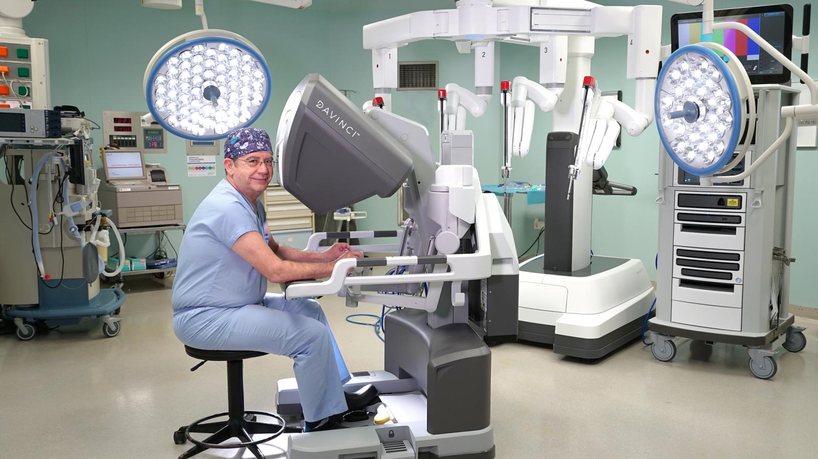
The surgeon operates the robot using a console to control the four robotic arms. The robot arms perform cutting, gripping, cauterizing, and retraction functions. The inside of the body is viewed in 3D through a camera and displayed on the console for the surgeon.
While the robot's arms are over the patient, a cardiac surgeon is present to hand over the necessary instruments to the robot's arms. The surgeon performing the surgery operates from a console located 3-4 meters away from the patient. The console's two arms are designed to control the four arms used on the patient.
One of the four arms on the patient is the camera, two of them move like the surgeon's right and left hands, while the fourth arm is used to move tissues that may obstruct the surgeon (retraction). In addition to being able to stabilize the arm the surgeon wants in any position, the surgeon can move the arms in ways that a human hand cannot, and eliminate the vibrations that a human hand would typically have.
The duration of surgeries performed with robotic surgery is becoming shorter with each passing day as experience increases.
Initially long, the duration of robotic heart surgeries has now equalized with the duration of traditional heart surgeries as experience is gained during the learning process. In fact, experienced hands in robotic surgery have even shortened this time compared to conventional surgeries. The reduction in surgical time has a very positive effect on the patient's recovery and return to life in surgeries where a heart-lung machine is used. In the future, with advancements in robotic technology and the growing experience of robotic surgeons, this time will continue to shorten.


